Adult Acquired Flatfoot Surgery: When Is It Needed?
Date:
Editor:
The Foot & Ankle Group
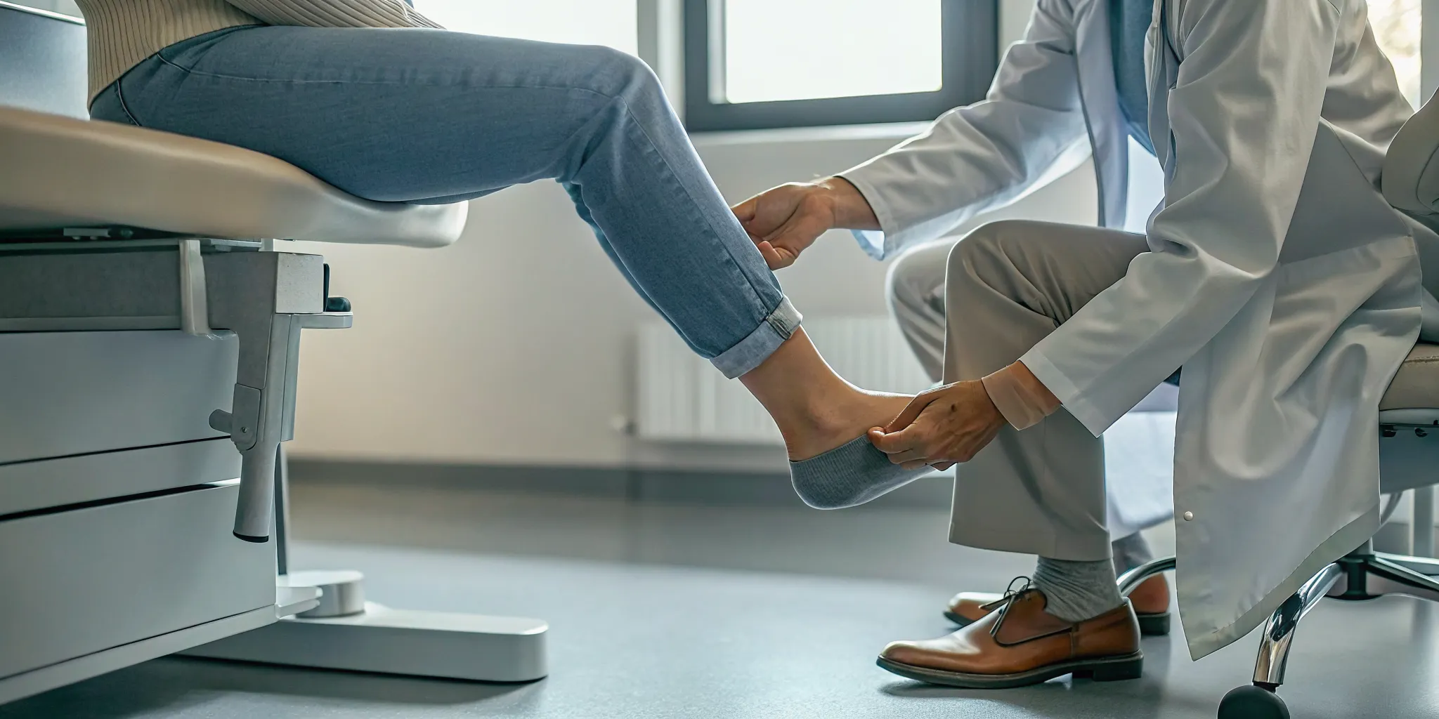
Flat feet are not always something a person has had since childhood. In some adults, the arch gradually lowers after the posterior tibial tendon, which helps support the arch and control the foot, becomes painful or weakened. You may notice inner-ankle soreness, swelling, a shoe that feels uneven, or difficulty standing on one foot.
Schedule a foot and ankle evaluation to find out whether adult acquired flatfoot surgery is the right next step for you, or whether conservative care should come first.
Adult acquired flatfoot surgery is considered when pain, weakness, or loss of function continues despite appropriate non-surgical care. But many people can first improve with support, immobilization, physical therapy, or bracing.
Because this condition can progress, an evaluation should look at more than the shape of the arch. Your foot and ankle specialist will assess tendon function, flexibility, alignment, symptoms, and how the problem affects walking. Understanding the underlying changes makes the stages and treatment choices easier to follow.
What Is Adult Acquired Flatfoot?
Adult acquired flatfoot deformity, often abbreviated AAFD, is a progressive change in which the foot's arch gradually loses its shape during adulthood. It is closely associated with posterior tibial tendon dysfunction, or PTTD. The condition is not simply a childhood foot type that has continued into later life. It develops when the structures that support the arch can no longer work effectively.
The posterior tibial tendon and the arch
The posterior tibial tendon runs along the inner side of the ankle and foot. It helps support the arch and assists with movements that stabilize the foot as you walk. When this tendon becomes irritated, stretched, degenerated, or otherwise insufficient, it may not provide the same support it once did. The arch can then begin to flatten, and the heel and ankle may gradually shift out of their normal alignment.
People may first notice arch pain, fatigue, swelling, or discomfort along the inside of the ankle. Symptoms can vary, and inner ankle pain does not always mean PTTD, but it is worth evaluating when it persists or affects walking. A foot and ankle specialist can assess the tendon, alignment, strength, flexibility, and degree of deformity rather than relying on symptoms alone.
Why adult acquired flatfoot can progress
Because the tendon and surrounding support system are involved, the change can be progressive if it is left unaddressed. As the arch lowers, other ligaments, joints, and tendons may take on additional stress. This does not mean every person with early symptoms will need surgery. Treatment is individualized, and many people begin with activity changes, supportive devices, physical therapy, or other nonsurgical care.
Medical literature increasingly uses the term progressive collapsing flatfoot deformity to describe this evolving condition. The newer name emphasizes that the foot can change over time, rather than suggesting that the issue is limited to a static flat-foot shape. If you are experiencing ongoing inner ankle pain or a noticeable change in your arch, an evaluation can clarify what is happening and which options are appropriate.
Sources: AAFD overview and current terminology review.
The Stages of Posterior Tibial Tendon Dysfunction
Posterior tibial tendon dysfunction (PTTD) usually develops gradually. The tendon runs behind the inside of the ankle and helps support the foot's arch. As it becomes painful or weakened, the arch may begin to collapse and the heel can shift outward. Because the condition is progressive, recognizing changes early can help guide treatment before the foot becomes more difficult to correct. Newer medical literature may call this condition progressive collapsing flatfoot deformity rather than adult acquired flatfoot deformity.
Stage I: Tendon pain without visible deformity. The earliest stage typically causes pain, tenderness, or swelling along the posterior tibial tendon behind the inner ankle. The foot's shape may still look normal, and the arch remains intact. Activities that place repeated stress on the tendon can make symptoms more noticeable.
Stage II: Flexible flatfoot. As the tendon loses its ability to support the arch, the arch begins to sag and the heel may shift outward. The foot is flatter, but the deformity is still flexible, meaning it can often be moved toward a more normal position. Patients may notice that shoes fit differently, the arch looks lower when standing, or walking becomes uncomfortable.
Stage III: Rigid flatfoot. Over time, the arch may become fixed in its collapsed position. The foot is no longer easily corrected by hand, and the hindfoot becomes stiff. Arthritis may develop in the joints around the middle and back of the foot, adding pain and limiting movement. Treatment at this stage must account for both the deformity and the joint changes.
Stage IV: Ankle involvement. In the most advanced stage, the progressive deformity extends to the ankle joint. The ankle may tilt inward as the supporting structures around the foot and ankle become affected. This stage can change alignment and stability beyond the foot itself, so evaluation should include the ankle as well as the arch.
These stages describe a general pattern, not a prediction of exactly how one person's condition will progress. A foot and ankle specialist can assess flexibility, alignment, tendon function, and joint health to determine the current stage and appropriate next step. Evaluation does not automatically mean adult acquired flatfoot surgery; many patients are first treated with supportive, non-surgical care.
Learn more about inner ankle conditions that may cause pain around the posterior tibial tendon.
Non-Surgical Treatments to Try First
Most people with adult-acquired flatfoot begin with a structured conservative plan rather than surgery. The right combination depends on how flexible the deformity is, how severe the pain has become, and whether the posterior tibial tendon is still functioning. In Our Expert Opinion, controlling a flare-up first makes it easier to restore strength and movement safely.
Conservative treatment options for adult-acquired flatfoot | ||
Treatment | What it does | Best for |
|---|---|---|
Orthotics | Supports the arch and uses a medial heel post to help correct heel angling. | Mild to moderate, flexible deformity after pain improves |
Physical therapy | Builds control through stretching, strengthening, and balance exercises. | Patients whose acute pain has subsided |
Bracing | Provides more substantial support and limits painful motion. | More developed deformity or inadequate shoe support |
Immobilization or CAM boot | Temporarily rests the tendon and reduces stress during a flare-up. | New or worsening pain and inflammation |
Anti-inflammatory medication | Helps reduce pain and inflammation as part of a broader plan. | Short-term symptom relief when medically appropriate |
A removable CAM walking boot is often used for three to six weeks during a painful flare-up. Once symptoms settle, an orthotic with arch support and a medial heel post may be fitted for mild to moderate deformity. More developed changes may require a short articulated ankle-foot orthosis or an Arizona brace.
Restoring strength after pain settles
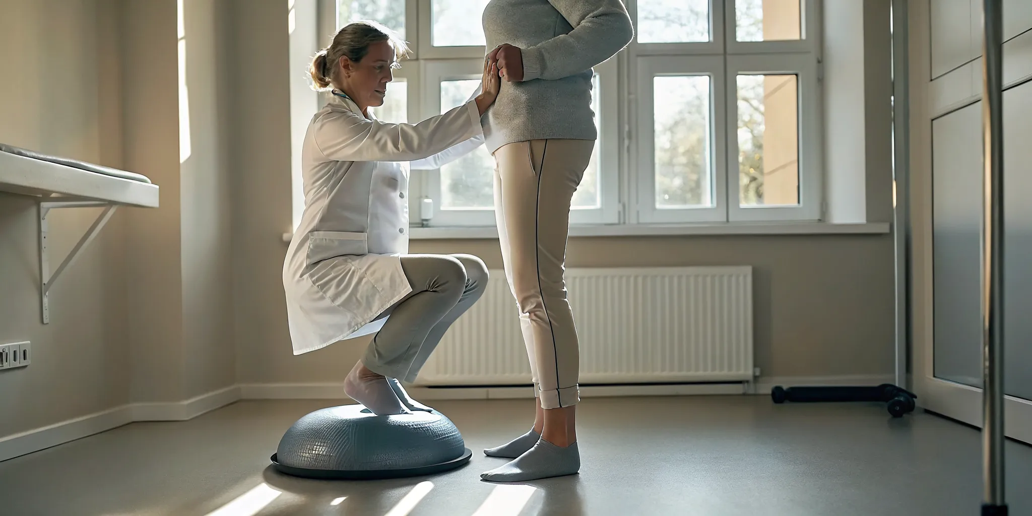
Physical therapy should progress at the appropriate time, not force an already irritated tendon to work harder. A program may include Achilles tendon stretching, inversion strengthening, toe flexor strengthening, and proprioception exercises to improve balance and foot control. Activity modification, supportive footwear, and weight management can also reduce repeated load on the arch. Anti-inflammatory medications, including NSAIDs, may help some patients, but a clinician should review whether they are safe for you.
For many patients, pain resolves with non-surgical care. If symptoms persist or function remains limited, a foot and ankle specialist can reassess the deformity and discuss the next step.
When Is Adult Acquired Flatfoot Surgery Necessary?
Surgery is usually considered when adult acquired flatfoot continues to cause meaningful pain that limits standing, work, or other daily activities after a thorough trial of non-surgical care. It may also become appropriate when the deformity is progressively collapsing and the changing alignment is making ordinary movement increasingly difficult.
Who may be a surgical candidate?
A candidate is not defined by the appearance of the arch alone. The decision depends on symptoms, physical examination, imaging, flexibility of the deformity, tendon and joint involvement, general health, and how much the condition affects the person's goals. A flexible deformity that remains painful despite appropriate treatment may be approached differently from a rigid deformity with arthritis or ankle involvement. A foot and ankle specialist can evaluate these factors and explain whether surgery is reasonable, premature, or unlikely to address the main source of discomfort.
In Our Expert Opinion, the purpose of an evaluation is not to push someone toward an operation. It is to identify why the arch is failing and match treatment to the individual. Research describes improvement with appropriately selected surgery when pain or functional limitations persist despite non-surgical treatment. Read the clinical review for additional background.
Conservative treatment comes first
Every stage of adult acquired flatfoot deformity should be given an opportunity to respond to non-surgical care before surgery is considered. Depending on the stage and symptoms, this may include temporary immobilization, a brace, supportive orthotics, activity changes, medication when appropriate, and physical therapy. Treatment may focus on reducing irritation, improving tendon and calf flexibility, strengthening supporting muscles, and making walking more comfortable. Pain can often improve without an operation, so persistence with a well-designed plan matters.
Setting realistic expectations
Modern surgical treatments for appropriately selected patients often exceed an 80 percent success rate, but that figure is not a promise or a guarantee for any individual. Recovery can be substantial, and risks, healing time, and the possibility of residual symptoms should be discussed openly. If surgery becomes part of the conversation, this overview of how flat feet are treated in adults may provide useful context before an evaluation.
What Flatfoot Reconstruction Involves
Flatfoot reconstruction is not one standard operation. It is a combination of procedures selected to address the specific position of your heel, arch, tendons, joints, and calf. The plan is based on your symptoms, physical examination, imaging, and how flexible or rigid the deformity is.
Realigning the heel and supporting the arch
A medializing calcaneal osteotomy realigns the heel bone so it shifts back underneath the lower leg. This improves the mechanics of the hindfoot and helps counter the forces that allow the arch to collapse. It also helps offload the spring ligament, an important structure that supports the arch, while increasing the foot's inversion moment.[1]
If the posterior tibial tendon is failing, a tendon transfer may be added. In many reconstructions, the flexor digitorum longus tendon is transferred to reinforce the weakened posterior tibial tendon. The goal is to provide more dependable support for the arch without treating the heel alignment in isolation.
Correcting the shape of the midfoot
Some patients need additional correction through the middle or outer portion of the foot. A lateral column lengthening uses a wedge graft to improve midfoot alignment and restore the relationship between the front and back of the foot. A Cotton osteotomy uses a wedge on the medial side of the midfoot to help bring the arch back into a more balanced position. These procedures are not required for every patient. They are chosen when the remaining deformity calls for them.
Addressing a tight calf
A tight calf can place extra stress on a reconstructed arch. Depending on the examination, the surgeon may recommend a gastrocnemius recession or, in selected cases, Achilles tendon lengthening. When the tightness is isolated to the gastrocnemius, recession is often preferred. It is less likely to over-lengthen the Achilles tendon and may allow a quicker recovery.[1]
In Our Expert Opinion, the purpose of combining these procedures is not simply to make the foot look straighter. It is to create a stable, plantigrade foot that can bear weight with less pain. To learn more about the conditions foot and ankle surgery can address, discuss your symptoms and treatment options with a foot and ankle specialist.
Recovery Timeline After Flatfoot Reconstruction
Recovery after reconstruction is gradual, and the pace depends on the procedures performed, your healing, and what follow-up imaging shows. A typical plan protects the reconstruction first, then adds motion, weight bearing, and strength in stages. Your surgeon may adjust this schedule if X-rays or your exam indicate that more time is needed.
Weeks 0-2: Protect the reconstruction. You will usually remain non-weight bearing in a splint for about two weeks. Keep the foot elevated as directed, use your crutches or another prescribed aid, and follow your surgical team's instructions for incision and swelling care. Range-of-motion exercises may begin around the two-week visit when your surgeon determines they are safe. The exact exercises depend on the reconstruction and should not be started independently.
Weeks 2-6: Transition to a short leg cast. After the initial splint period, many patients move into a short leg cast for approximately four weeks. Non-weight bearing commonly continues during this phase. In total, non-weight bearing may last roughly six to 10 weeks, particularly when several procedures are combined. This is one reason to plan ahead for work, transportation, and help at home.
Around weeks 6-10: Let imaging guide weight bearing. Follow-up X-rays and your clinical examination help determine when it is appropriate to begin putting weight on the foot. If healing is progressing as expected, weight bearing is introduced gradually rather than all at once. You may transition from the cast into a walking boot while increasing activity under specific instructions.
Around week 6 and beyond: Begin physical therapy. Physical therapy often starts near six weeks, although the timing varies by procedure and healing. Early therapy focuses on restoring motion, reducing stiffness, and rebuilding a safe walking pattern. Your therapist can then help address strength, balance, and gait mechanics as your restrictions change.
Around week 12 and later: Add progressive strengthening. Progressive strengthening commonly begins around 12 weeks after surgery. Exercises advance in stages, based on pain, swelling, strength, gait, and imaging rather than a calendar alone. Returning to regular shoes and higher-impact activities may take additional time.
Weeks 8-12 through the following months: Add support as needed. An orthotic or shoe insert is often considered around eight to 12 weeks to support the foot during recovery. Some patients use this support for six to 12 months, depending on symptoms, alignment, and activity demands. Your surgeon or therapist will advise whether an insert is appropriate and when to change it.
It is normal for recovery to feel uneven, with swelling or fatigue increasing after a change in activity. Contact your surgical team if symptoms are worsening or you are unsure whether a new activity is appropriate. A carefully paced flatfoot reconstruction recovery gives healing tissues time to adapt while your care team uses imaging and examination findings to guide each next step.
Published clinical guidance describes non-weight bearing followed by gradual progression, with range-of-motion exercises around two weeks and strengthening around 12 weeks, but your individualized plan remains the controlling recommendation.
Ready to talk through your flatfoot symptoms? Contact The Foot and Ankle Group to schedule an evaluation and learn whether conservative care or reconstruction fits your situation.
Frequently Asked Questions
Can flat feet be corrected in adults?
Often, symptoms and function can improve without surgery, especially when the deformity is flexible. Treatment may include activity changes, a removable walking boot, supportive orthotics, bracing, medication, and physical therapy. An orthotic with arch support and a medial heel post may help manage mild to moderate deformity. A rigid or progressing deformity may require a different plan, so an evaluation is important before choosing treatment.
How long does it take to walk after flatfoot surgery?
The timeline depends on the procedures performed and how healing progresses. A common protocol uses about two weeks without weight on the foot in a splint, followed by four or more weeks in a removable cast. Patients may then gradually progress to full weight bearing over approximately four weeks. Range-of-motion exercises may begin around two weeks, while strengthening commonly starts near 12 weeks. These are general guidelines, not a personal recovery promise. Source: clinical review of adult-acquired flatfoot treatment.
How do you fix an adult-acquired flatfoot?
The first step is identifying the cause, flexibility, stage, and effect on daily activity. Early treatment usually focuses on reducing tendon irritation and supporting the arch. If pain or functional limits continue despite nonsurgical care, reconstruction may combine procedures such as a heel bone adjustment, tendon transfer, calf muscle lengthening, or other bone corrections. The appropriate combination is individualized rather than selected from a single standard operation.
How painful is flatfoot surgery?
Some pain, swelling, and stiffness are expected after reconstruction, particularly during the first several days and while the foot is protected from weight. Your surgeon will provide a pain-control plan and instructions for elevation, medication, wound care, and warning signs. Pain that is severe, worsening, or not controlled as expected should be reported promptly. In Our Expert Opinion, discussing pain expectations before surgery can make the recovery plan clearer.
Schedule a Foot and Ankle Evaluation
An evaluation can clarify whether your adult-acquired flatfoot symptoms may respond to conservative care or whether reconstruction should be considered. The Foot and Ankle Group can review your symptoms, examine your foot and ankle, and discuss reasonable next steps based on your needs. Reach our team at (609) 291-0960 or schedule your foot and ankle evaluation online to get started.